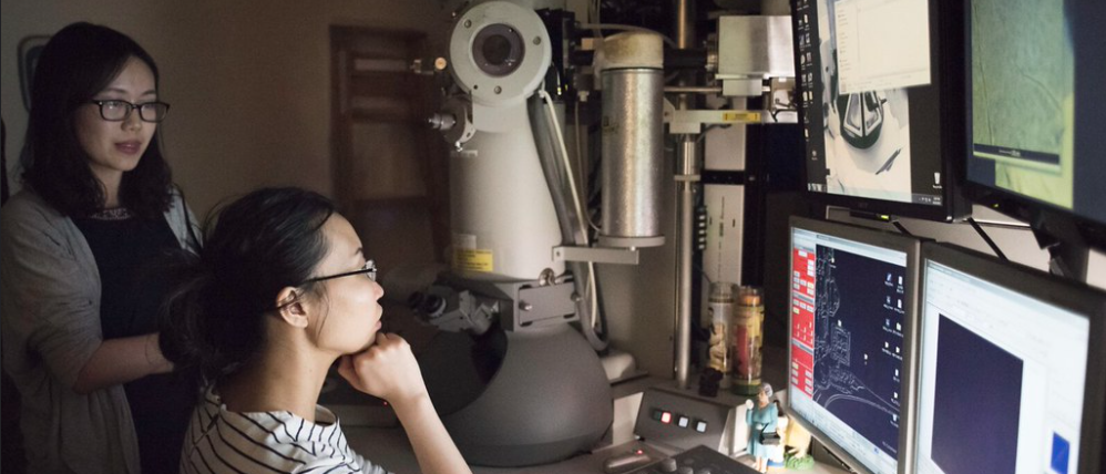
Electron Microscopy Facility
at Cornell University
PARADIM’s electron microscopy (EM) facility leverages and expands on existing capabilities in the Cornell Center for Materials Research (CCMR, a research center that includes an NSF-funded Materials Research Science and Engineering Center, MRSEC DMR-1719875) giving users access to a unique set of advanced analysis tools for materials characterization. These electron microscopy facilities are part of the Cornell Center for Materials Research but can be accessed via the PARADIM user program. Reviewed and approved PARADIM user projects receive access to the facilities and to imaging and sample preparation support from PARADIM staff. As with other PARADIM resources, access is without charge to approved US academic and government projects. Preference will be given to projects in which characterization is part of larger materials by design effort.
Capabilities and Highlights
Electron Microscopy New Capabilities
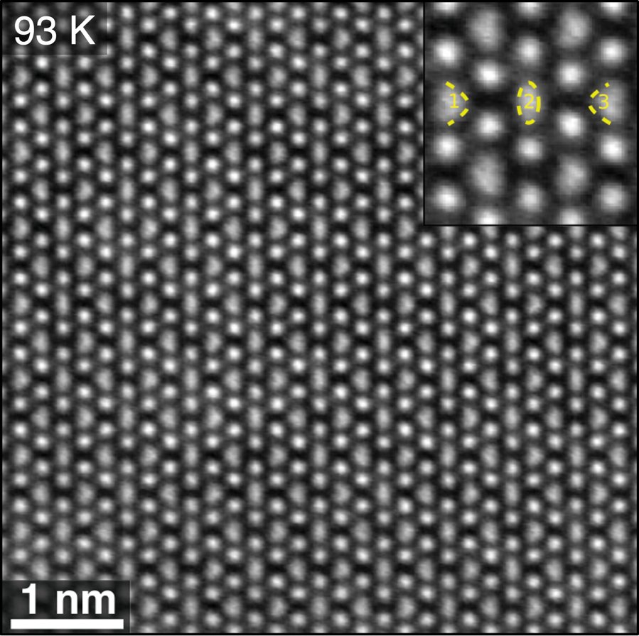
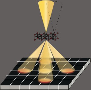
Sub-Angstrom STEM Imaging
PARADIM offers sub-Angstrom imaging of materials at room and cryogenic temperatures.
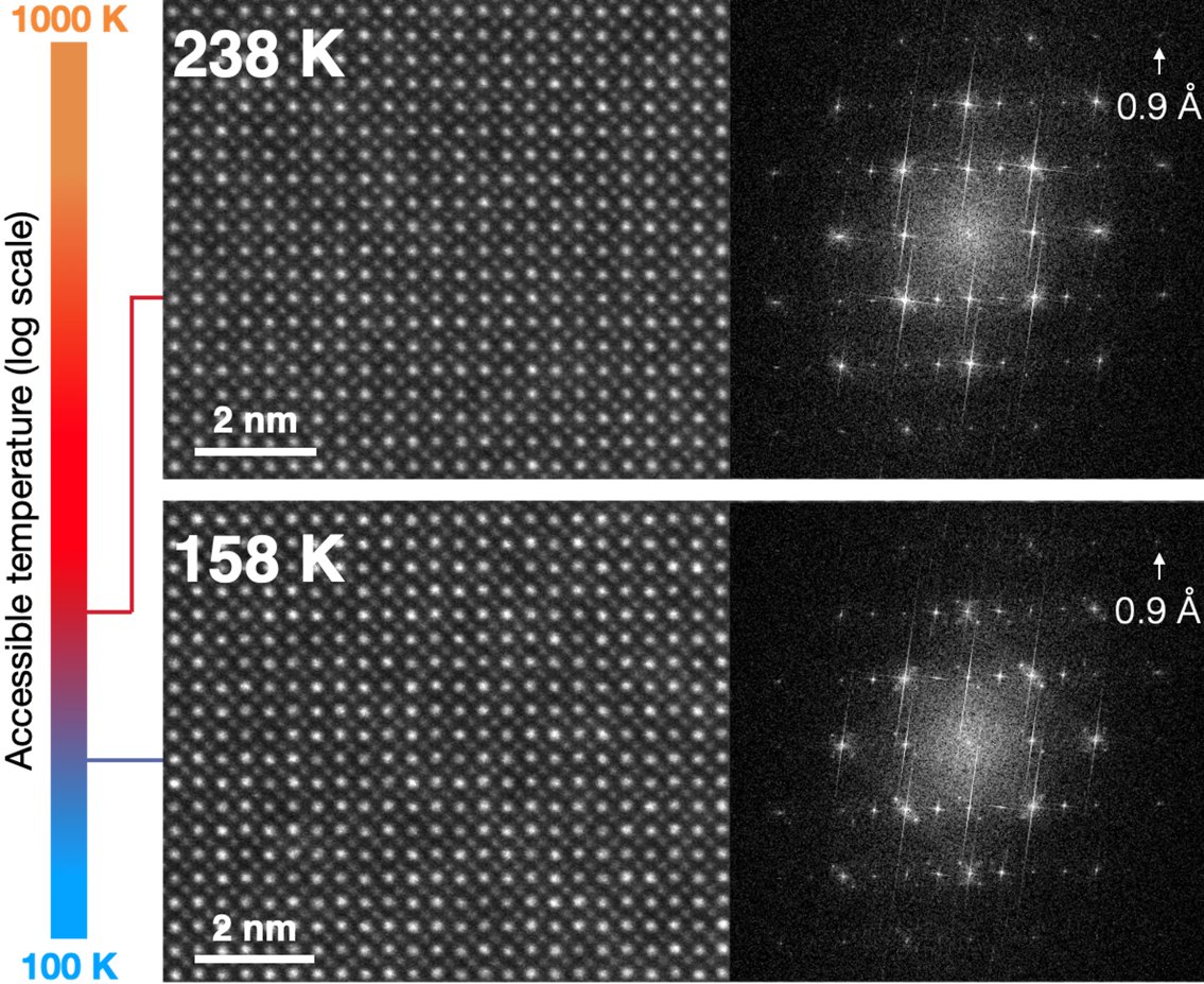
Variable temperature cryo-STEM
A new sample holder enables atomic-resolution experiments at variable cryogenic temperatures for tracking of phase transitions.
Electron Ptychography
E-ptychography offers never before seen resolution.
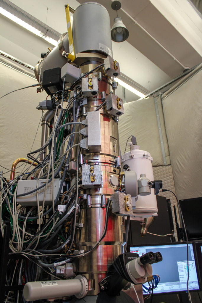
Tools

TFS Spectra 300 Andromeda S/TEM + X-CFEG
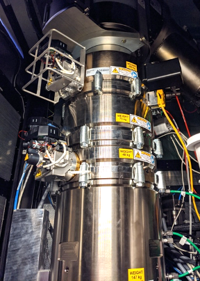
TFS Spectra 300 Kraken S/TEM + X-CFEG
FEI Titan Themis 300
EMPAD

Tescan Amber X 2 Xenon Plasma FIB - Fall 2025
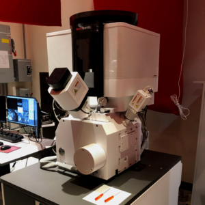
TFS Helios G4 UX DualBeam FIB
Electron Microscopy Team
Dr. Steven Zeltmann
Steven is an electron microscopist specializing in using 4D-STEM to solve materials science problems.

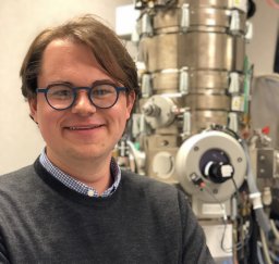
Dr. David Muller
Leading the PARADIM Electron Microscopy Facility, Dr. David Muller continues to develop groundbreaking capabilities to support users. With electron ptychography and the new EMPAD detector his group has achieved imaging at record-breaking resolution.